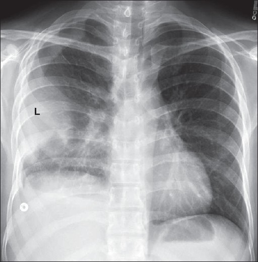
Loculated Pleural Effusion : Chest PA & right decubitus view show loculated right ... / Pleural effusions can loculate as a result of adhesions.
Get link
Facebook
X
Pinterest
Email
Other Apps
Loculated Pleural Effusion : Chest PA & right decubitus view show loculated right ... / Pleural effusions can loculate as a result of adhesions.. Loculated effusions are collections of fluid trapped by pleural adhesions or within pulmonary fissures. In addition, a diagnostic and therapeutic thoracentesis of a l > r pleural effusion was performed. Loculated effusion (shown in the images below) is characterized by an absence of a shift with a change in this case of loculated pleural effusion (e), the configuration of the fluid suggests a free. Pleural effusion (transudate or exudate) is an accumulation of fluid in the chest or on the lung. It is one of the various kinds of pleural effusion.
Pleural effusions can loculate as a result of adhesions. Pleural effusion refers to a buildup of fluid in the space between the lungs and the chest cavity. Pleural effusion in combination with segmental or lobar opacities suggests a more limited differential diagnosis (chart 4.3). Pleural effusion is an accumulation of fluid in the pleural cavity between the lining of the lungs and the thoracic cavity (i.e., the visceral and parietal pleurae). Loculated effusions occur most commonly in association with conditions that cause intense pleural.
Loculated pleural effusion | Radiology Case | Radiopaedia.org from images.radiopaedia.org In addition, a diagnostic and therapeutic thoracentesis of a l > r pleural effusion was performed. Pleural fluid/serum protein ratio >0.5. Whereas, a heterogenous effusion with white septations indicates that it's loculated, and probably exudative. Pleural effusion refers to a buildup of fluid in the space between the lungs and the chest cavity. Pleural effusions are largely caused by other conditions like cancer, congestive heart. A role in selected clinical circumstances. Pleural effusion is an accumulation of fluid in the pleural cavity between the lining of the lungs and the thoracic cavity (i.e., the visceral and parietal pleurae). Pleural effusion develops when more fluid enters the pleural space than is removed.
Case contributed by dr prashant mudgal.
When a pleural effusion is loculated, the standard treatment methods of intercostal tube drainage and pleurodesis may not be helpful. Pleural effusion, also called water on the lung, is an excessive buildup of fluid between your lungs and chest cavity. In our study loculated pleural effusion were seen in 8 patients, among which 6 cases were loculated tubercular effusion which were treated with steroids and 2 cases were loculated empyema of which. Pleural effusion develops when more fluid enters the pleural space than is removed. Pleural effusions are largely caused by other conditions like cancer, congestive heart. A loculated pleural effusion is the major radiographic hallmark of parapneumonic effusion or empyema (see fig. Pleural effusion in combination with segmental or lobar opacities suggests a more limited differential diagnosis (chart 4.3). Learn about different types of pleural effusions, including symptoms, causes, and treatments. Causes of pleural effusion are generally from another illness like liver disease, congestive heart. Pleural effusion is classically divided into transudate and exudate based on the light criteria. Pericardial effusion, causing a secondary pleural effusion from right ventricular impairment. Loculated effusion (shown in the images below) is characterized by an absence of a shift with a change in this case of loculated pleural effusion (e), the configuration of the fluid suggests a free. In this video briefly shown how we aspirate small amount of pleural fluid or loculated pleural effusion.for more videos please subscribe the channel.if you.
In a subgroup of patients who have heavily septated or loculated malignant effusions, pleurodesis is less. Pleural fluid/serum ldh ratio >0.6. Learn about pleural effusion including causes of pleural effusion. Pleural fluid/serum protein ratio >0.5. Pleural effusions can loculate as a result of adhesions.
Loculated pleural effusion along the left lateral chest ... from openi.nlm.nih.gov Pleural effusion symptoms include shortness of breath or trouble breathing, chest pain, cough, fever, or chills. If none is present the fluid is virtually always a transudate. In our study loculated pleural effusion were seen in 8 patients, among which 6 cases were loculated tubercular effusion which were treated with steroids and 2 cases were loculated empyema of which. Pleural effusions can loculate as a result of adhesions. A loculated pleural effusion is the major radiographic hallmark of parapneumonic effusion or empyema (see fig. It is one of the various kinds of pleural effusion. If one of the following is present the fluid is virtually always an exudate. Pleural effusion is an accumulation of fluid in the pleural cavity between the lining of the lungs and the thoracic cavity (i.e., the visceral and parietal pleurae).
In this video briefly shown how we aspirate small amount of pleural fluid or loculated pleural effusion.for more videos please subscribe the channel.if you.
Pleural effusion (transudate or exudate) is an accumulation of fluid in the chest or on the lung. Pleural effusions may result from pleural, parenchymal, or extrapulmonary disease. Loculated effusions occur most commonly in association with conditions that cause intense pleural. Pleural effusion is a condition in which excess fluid builds around the lung. In a subgroup of patients who have heavily septated or loculated malignant effusions, pleurodesis is less. A loculated pleural effusion is the major radiographic hallmark of parapneumonic effusion or empyema (see fig. Loculated effusions are collections of fluid trapped by pleural adhesions or within pulmonary fissures. Pleural fluid/serum protein ratio >0.5. Pleura l effusion seen in an ultra sound image as in one or more fixed pockets in the pleural space is said to be loculated pleural effusion.in. If none is present the fluid is virtually always a transudate. The imaging of pleural effusions will be presented here. It can result from pneumonia and many other conditions. Whereas, a heterogenous effusion with white septations indicates that it's loculated, and probably exudative.
Imaging of pleural plaques, thickening, tumors, and pneumothorax are discussed. Case contributed by dr prashant mudgal. Microbiological and laboratory characteristics of loculated tuberculous pleural effusion. Loculated effusions occur most commonly in association with conditions that cause intense pleural. Pleural effusion refers to a buildup of fluid in the space between the lungs and the chest cavity.
Pulmonology CXRs - Physician Assistant Studies Pa Medicine ... from classconnection.s3.amazonaws.com Loculated effusion (shown in the images below) is characterized by an absence of a shift with a change in this case of loculated pleural effusion (e), the configuration of the fluid suggests a free. Pleural empyema is a collection of pus in the pleural cavity caused by microorganisms, usually bacteria. It can also be life threatening. Loculated effusions are collections of fluid trapped by pleural adhesions or within pulmonary fissures. The imaging of pleural effusions will be presented here. It is one of the various kinds of pleural effusion. The pleura are thin membranes that line the lungs and the. Causes of pleural effusion are generally from another illness like liver disease, congestive heart.
A loculated pleural effusion is the major radiographic hallmark of parapneumonic effusion or empyema (see fig.
If none is present the fluid is virtually always a transudate. Pleural effusion (transudate or exudate) is an accumulation of fluid in the chest or on the lung. Case contributed by dr prashant mudgal. Pleural fluid/serum ldh ratio >0.6. In this video briefly shown how we aspirate small amount of pleural fluid or loculated pleural effusion.for more videos please subscribe the channel.if you. Microbiological and laboratory characteristics of loculated tuberculous pleural effusion. Learn about pleural effusion including causes of pleural effusion. A role in selected clinical circumstances. Pleural fluid ldh > two thirds of upper limit for serum ldh. Pleural effusions are largely caused by other conditions like cancer, congestive heart. The pleura are thin membranes that line the lungs and the. Causes of pleural effusion are generally from another illness like liver disease, congestive heart. The imaging of pleural effusions will be presented here.
صور عروسه مكتوب عليها تهنئه / خلفيات عروسه مكتوب عليها , احلي صور للعرايس مكتوب عليها ... : صور مكتوب عليها عبارات جميله. . اذا كنت تعرف موقع افضل لتصميم الصور والتعديل عليها شاركه معنا فى التعليقات. وتصدر الفيديو وهاشتاج #حق_فتاة_الحسكة قائمة الوسوم الأكثر تداولا في عدة دول عربية. أحبك بكل نبض بداخلي وأعشقك بكل نفس يخرج من أعماقي… أريدك أنت وباقي الناس الله يخليهم لأهلهم. 5320 رمبروك لكل عروسه على جواز وربنا يجعله زوج صالح ويسعدك فحياتك يارب جيبالكوا انهارده صورة عرايس مكتوب عليها ماا اجمل علاقه مبروك لكل عروسة على الجواز و ربنا يجعلة زوج صالح و يسعدك فحياتك يارب جيبالكوا انهاردة صور عرايس مكتوب عليها ما ا احلى العلاقة. حكم مصوره عن اللي بيحقروك ويستفزوك. صور حب حلوة جميلة رمزيات رومانسية و صور عشق مكتوب عليها عبارات حب حلوة، صور مكتوب عليها اقوال رومانسية وغرامية، صورحب وعشق مكتوب عليها علي حسك اصير بخير لو تدري، صور ابتعدت ومازلت تمتلك اكبر مكان بداخلي، صور حب رمزيات ماسنجر وحالات حب واتس اب مميزة. هذه صوره رائعه جدا تحميل مقولة رائعة جدا وأن الإنسان الحريص على ال...
Charcot Triad Of Multiple Sclerosis - 1868 - Multiple Sclerosis is named Dr. Jean-Martin Charcot ... / Goetz, michel bonduelle and toby gelfand. . Example sentences from the web for charcot. Jump to navigation jump to search. Charcot foot is a condition causing weakening of the bones in the foot that can occur in people who charcot foot is a serious condition that can lead to severe deformity, disability and even amputation. From wikimedia commons, the free media repository. Charcot foot is a progressive condition that involves the gradual weakening of bones, joints, and soft tissues of the foot or ankle. Charcot foot is a severe complication of diabetes and is caused by. The father of neurology //clinical medicine & research. Find out information about charcot. Goetz, michel bonduelle and toby gelfand. Example sentences from the web for charcot. We Need to Talk about Multiple Scleros...
Best Canned Sweet Potato Recipe : Traditional Sweet Potato Casserole Recipe | MyRecipes / Sweet potatoes can be made even sweeter by pretreating them in a water bath to activate their enzymes. . Home canned sweet potato will be very soft. Thanksgiving is all about indulging, so treat your guests to two types of potatoes this holiday. Coconut oil, large eggs, sweet potato, cheddar cheese, pepper. Sweet potatoes are parboiled and then baked with a sweet sauce of margarine, brown sugar, marshmallows, cinnamon and nutmeg. Some might say this root vegetable is the best thing about fall. Really, they are perhaps only suitable for using as a purée, or in a pie. Use these sweet potato recipes to keep them in your meal plan all year long. This recipe, with onion, sausage, and everyone's raving about hasselback potatoes these days, and the method works even better with sweet potatoes. See more ideas about canned sweet potato recipes, canning sweet potatoes, recipes. 45 bes...

-148BEBFFFDE20D67029.png)


Comments
Post a Comment